Art Exhibtions
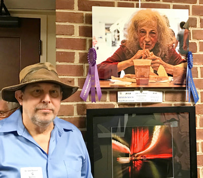
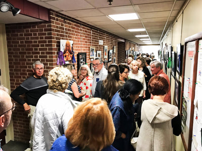
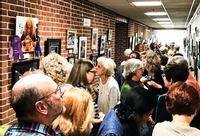
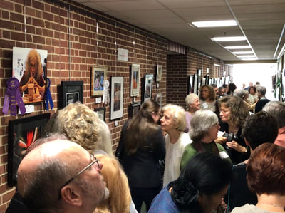
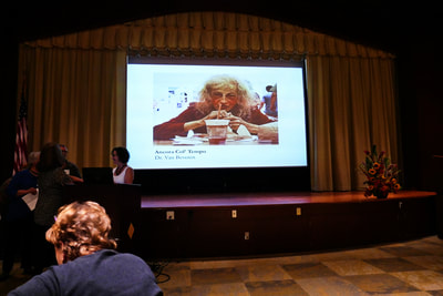
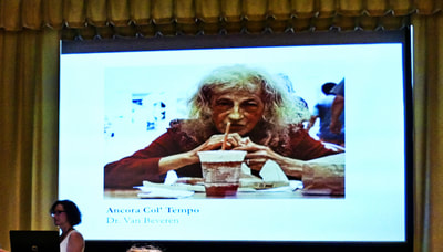
"Hey That's Me" Photografee
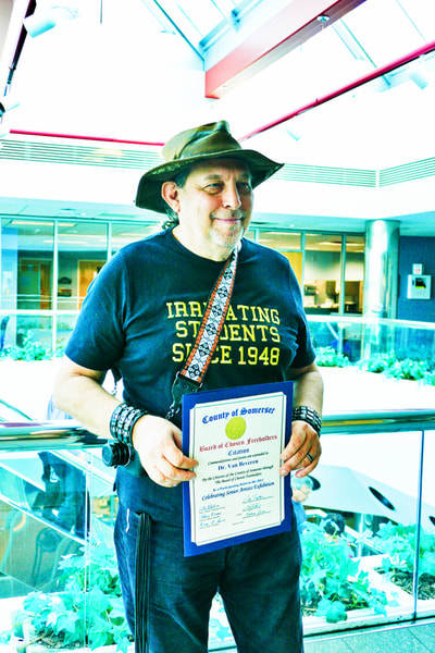
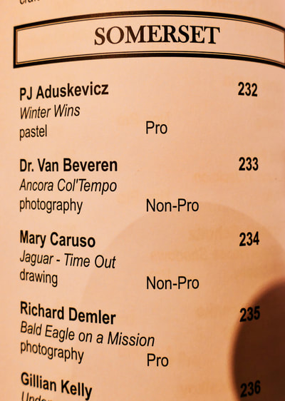
Explore the world through the lens of Dr. Van Beveren during his worldwide travels. His photographs are available for showing and purchasing. Each photo can be printed on a strong metallic frame or canvas, and are available in the following sizes:
Shipping and Handling will be calculated, depending on your zip code. Quality Guaranteed. Some photos are not available in all sizes.
Street Art
Microscopic Art
High-resolution images printed on strong metallic frames, which can be mounted in your place of business or home with relative ease. Request any size frame from 11" x 14" - 12" x 12" - 16" x 20" - 20" x 20" - 20" x 30" - 24" x 36".
One of the advantages of this type of art is that it can be hung horizontally or vertically and no one will be the wiser!
None of these photographs have seen the inside of a photo-shop. These photos were all taken with an Olympus BH2 Microscope, a Nomarski condenser and a minimum exaggeration of 400X - but some may be much higher. Colors have been manipulated by both liquid dyes (such as India ink, oil, vinegars, etc.) and several kinds of microscopic filters. Attached to the microscope was a small-bodied Olympus camera with a M. Zuiko lens, aperture @ 45mm. and an f1.8 stop.
The subject of these photos is the human body. All of the original pictures were taken from my patients' liquids and organ secretions and I have fun listening to viewers guess and palaver as they contemplate their origins. None of the photographs are identifiable or have been used in diagnosis.
I hope you will enjoy looking at them as much as I did photographing them.
Price list available upon request.
Dr. Van Beveren, PhD
Nutritional Biochemist & Clinical Physiologist
Princeton Health Integration Center
609-924-7337
